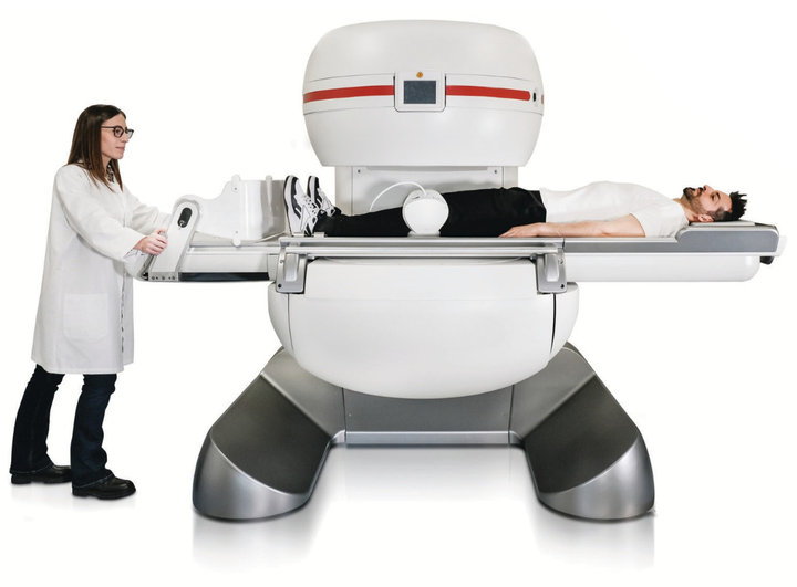
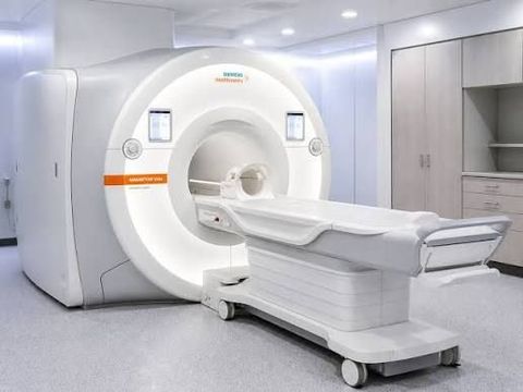
MRI is particularly valuable for imaging soft tissues, the brain, spinal cord, joints, muscles, abdominal organs, blood vessels, and other anatomical structures. Unlike X-ray and CT, MRI does not use ionizing radiation.
Today's MRI systems are evolving beyond traditional enclosed scanners. Developments include wide-bore designs, higher-field systems, low-field and portable MRI, faster imaging sequences, artificial intelligence-assisted reconstruction, quantitative imaging, and increasingly automated workflows.
What Is a Modern MRI Machine?
An MRI machine is a medical imaging system that uses a magnetic field and radiofrequency energy to generate signals from hydrogen protons within the body. Computers process these signals into detailed cross-sectional and three-dimensional images.
A modern MRI system generally includes:
-
Main magnet
-
Gradient coils
-
Radiofrequency coils
-
Patient table
-
RF electronics
-
Computer and reconstruction hardware
-
Imaging software
-
Cooling infrastructure
-
Safety and monitoring systems
The scanner operates as part of a larger MRI suite rather than as an isolated piece of equipment.
How Modern MRI Technology Works
The human body contains large amounts of hydrogen, particularly in water and fat. The MRI's magnetic field influences the orientation of hydrogen protons.
Radiofrequency energy then temporarily changes their state. As the protons return toward equilibrium, they produce signals that can be detected by the scanner.
The system combines these signals with spatial information generated by gradient fields to construct images.
This fundamental process enables MRI to create highly detailed anatomical images without using ionizing radiation.
Major Types of Modern MRI Machines
Closed-Bore MRI
Closed-bore MRI systems use a cylindrical magnet with a tunnel-like opening.
They remain among the most widely used configurations because they can provide strong imaging performance across a broad range of clinical applications.
Wide-Bore MRI
Wide-bore systems provide a larger opening than conventional systems.
Potential advantages include:
-
Improved patient accessibility
-
Greater physical comfort
-
Easier positioning
-
Better accommodation for some patients
Wide-bore designs have also become an important area of development for higher-field MRI systems.
Open MRI
Open MRI systems use a more open physical configuration.
They can be useful where patient accessibility and comfort are important. Open systems may be particularly relevant for some patients who have difficulty with conventional enclosed scanners.
However, MRI configurations differ considerably in magnetic-field strength and imaging capabilities, so "open" does not by itself describe overall system performance.
Low-Field MRI
Low-field MRI operates at substantially lower magnetic-field strengths than conventional hospital systems.
Recent developments have made some low-field systems smaller and more mobile. NIST notes that certain newer low-field systems can potentially be transported to locations such as intensive-care environments, although these technologies are still developing and have limitations compared with conventional MRI.
1.5 Tesla MRI
1.5T systems are widely used in clinical imaging.
They provide a balance between imaging capability, established clinical applications, infrastructure requirements, and operational considerations.
Typical applications include:
-
Neurological imaging
-
Spine imaging
-
Musculoskeletal examinations
-
Abdominal imaging
-
Pelvic imaging
-
Cardiac imaging
-
General diagnostic MRI
3 Tesla MRI
3T systems use a stronger magnetic field than 1.5T systems.
They can provide higher signal-to-noise characteristics for many applications and can support high-resolution imaging and advanced techniques.
3T MRI has become particularly important in advanced clinical imaging and research environments.
7 Tesla MRI
Ultra-high-field MRI systems such as 7T systems operate at substantially higher field strength.
They are used primarily in specialized clinical and research environments and can provide opportunities for highly detailed imaging.
Their use requires specialized technical, safety, and operational considerations.
MRI Field Strength Explained
MRI field strength is measured in tesla (T).
Common categories include:
-
Low-field systems
-
1.5T systems
-
3T systems
-
Ultra-high-field systems such as 7T
Higher field strength can provide advantages in signal characteristics and certain imaging applications, but it does not automatically mean that every examination will be superior.
System selection depends on:
-
Clinical applications
-
Patient population
-
Imaging protocols
-
Infrastructure
-
Workflow
-
Safety requirements
-
Available technical expertise
Important MRI Technologies
Gradient Technology
Gradient coils create controlled variations in the magnetic field.
Gradient performance influences:
-
Spatial encoding
-
Imaging speed
-
Resolution
-
Diffusion imaging
-
Advanced imaging sequences
RF Technology
Radiofrequency systems transmit energy and receive signals from the body.
Specialized RF coils are designed for different anatomical areas, including:
-
Head
-
Spine
-
Knee
-
Shoulder
-
Breast
-
Abdomen
-
Extremities
Parallel Imaging
Parallel imaging uses multiple RF coil elements to accelerate data acquisition.
This can help reduce examination times while maintaining useful image quality.
Compressed Sensing
Compressed-sensing techniques can reconstruct images from fewer measurements under appropriate conditions.
They can contribute to faster MRI examinations.
Simultaneous Multi-Slice Imaging
Some advanced techniques allow multiple imaging sections to be acquired simultaneously, helping accelerate certain MRI protocols.
AI in Modern MRI
Artificial intelligence is becoming increasingly important in MRI technology.
Potential applications include:
-
Image reconstruction
-
Noise reduction
-
Scan acceleration
-
Automated positioning
-
Protocol assistance
-
Image segmentation
-
Quality assessment
-
Workflow optimization
Recent industry discussions have highlighted AI-assisted automation across MRI workflows, including scan planning, acquisition, and quality control.
AI can therefore contribute not only to image processing but also to the broader imaging workflow.
Quantitative MRI
Traditional MRI primarily provides detailed anatomical images.
Quantitative MRI attempts to generate measurable tissue parameters.
Examples include:
-
T1 mapping
-
T2 mapping
-
Relaxometry
-
Diffusion measurements
-
Perfusion measurements
NIST notes that quantitative MRI is already being explored for specialized applications, including liver-related imaging, while research continues into improving measurement accuracy.
Functional MRI
Functional MRI, or fMRI, measures changes associated with brain activity.
It can be used in:
-
Neuroscience research
-
Brain-function studies
-
Pre-surgical mapping
-
Cognitive research
It provides information that complements conventional anatomical MRI.
Diffusion MRI
Diffusion-weighted imaging evaluates the movement of water molecules within tissue.
It has important applications in neurological imaging and can provide information about tissue characteristics that may not be visible through conventional structural imaging alone.
MR Angiography
Magnetic resonance angiography, or MRA, is used to visualize blood vessels.
Depending on the examination and clinical objective, MRA can be performed with or without contrast agents.
Cardiac MRI
Cardiac MRI provides detailed information about the structure and function of the heart.
It can be used to evaluate:
-
Cardiac anatomy
-
Ventricular function
-
Myocardial characteristics
-
Blood flow
-
Certain cardiovascular conditions
Specialized cardiac sequences and synchronization techniques are used for these examinations.
Musculoskeletal MRI
MRI is particularly useful for soft-tissue imaging.
Musculoskeletal applications include examination of:
-
Knees
-
Shoulders
-
Hips
-
Wrists
-
Ankles
-
Muscles
-
Tendons
-
Ligaments
-
Cartilage
MRI can provide detailed information about structures that may be difficult to evaluate with some other imaging methods.
Neurological Applications
MRI plays an important role in neurological imaging.
Applications include imaging of:
-
Brain
-
Spinal cord
-
Cerebral blood vessels
-
Cranial nerves
-
Brain tumors
-
Stroke-related changes
-
Degenerative neurological conditions
MRI's ability to provide detailed soft-tissue contrast makes it particularly valuable in this field.
Oncology Applications
MRI can contribute to cancer imaging and treatment monitoring.
It may be used for evaluating:
-
Tumor location
-
Tissue characteristics
-
Disease extent
-
Treatment response
The specific MRI protocol depends on the anatomical region and clinical question.
MRI Installation Requirements
Installing a modern MRI machine requires detailed facility planning.
Major considerations can include:
-
MRI room dimensions
-
Magnet delivery route
-
Structural requirements
-
RF shielding
-
Magnetic-field management
-
Electrical infrastructure
-
HVAC
-
Cooling
-
Equipment-room requirements
-
Patient preparation areas
-
Safety zones
The exact requirements depend on the MRI system and facility.
MRI Room Planning
A typical MRI suite can contain:
-
Scan room
-
Control room
-
Equipment room
-
Patient preparation area
-
Changing facilities
-
Supporting clinical spaces
The layout should support safe patient movement and controlled access to the MRI environment.
RF Shielding
Radiofrequency shielding helps protect MRI signal acquisition from external electromagnetic interference.
A properly designed shielded environment can reduce unwanted RF interference and associated image artifacts.
Magnetic-Field Safety
The MRI magnetic field creates safety considerations that differ from many other medical imaging technologies.
Ferromagnetic objects can become dangerous projectiles when brought near a strong MRI magnet.
MRI facilities therefore use controlled access areas and screening procedures.
MRI Screening
Patients and staff may need to be screened for:
-
Implanted medical devices
-
Metallic foreign bodies
-
Certain surgical implants
-
Ferromagnetic objects
-
Other MRI-related risks
The MRI compatibility status of an implant should be verified according to its specific labeling and conditions.
Some devices are classified as MR Conditional, meaning they can be scanned only under defined conditions.
Electrical and Cooling Infrastructure
Modern MRI systems require appropriate facility infrastructure.
This may include:
-
Stable electrical supply
-
Grounding
-
Power conditioning
-
HVAC
-
Equipment-room cooling
-
Chilled-water infrastructure where applicable
Exact technical specifications should be established during the site-planning process based on the selected MRI system.
Quench Planning
Superconducting MRI systems can require specific planning for a potential quench event.
A quench involves rapid loss of superconductivity and can cause stored cryogenic material to transition rapidly into gas.
Depending on the system, the facility may require:
-
Quench ventilation
-
Pressure-relief arrangements
-
Appropriate room design
-
Emergency procedures
These requirements should be addressed during facility planning.
MRI Patient Comfort Technology
Modern MRI manufacturers increasingly focus on patient experience.
Features can include:
-
Wide-bore configurations
-
Improved lighting
-
Audio systems
-
Noise-management technology
-
Patient communication systems
-
Faster scanning
-
Improved ventilation
-
Visual distractions
These features can help make the examination environment more comfortable.
MRI Applications Across Healthcare
Modern MRI technology is used across numerous medical specialties.
| Healthcare Area | Common MRI Applications |
|---|
| Neurology | Brain and spinal imaging |
| Orthopedics | Joints, muscles, ligaments and cartilage |
| Cardiology | Heart structure and function |
| Oncology | Tumor imaging and monitoring |
| Gastroenterology | Abdominal and pelvic imaging |
| Vascular Imaging | Blood-vessel evaluation |
| Pediatrics | Selected soft-tissue examinations |
| Research | Functional and quantitative imaging |
MRI is used for disease detection, diagnosis, and treatment monitoring across many clinical areas.
Mobile and Portable MRI
One of the most notable developments is the emergence of smaller MRI systems.
Traditional MRI scanners are large and generally located in hospitals or dedicated imaging facilities. New low-field technologies are exploring more mobile configurations.
Potential applications include:
-
Intensive-care environments
-
Smaller healthcare facilities
-
Emergency settings
-
Remote locations
-
Specialized neurological imaging
NIST reports that some emerging low-field MRI systems are small enough to be transported to patients, although their image capabilities and clinical applications differ from conventional hospital MRI.
MRI and Hospital Digital Infrastructure
Modern MRI systems generate substantial quantities of imaging data.
Digital integration commonly involves:
-
PACS
-
RIS
-
DICOM
-
Hospital information systems
-
Electronic medical records
-
Secure imaging archives
Efficient integration helps imaging departments manage, transfer, store, and review MRI examinations.
MRI Workflow Automation
Automation can influence several stages of the imaging workflow.
Potential areas include:
-
Patient positioning
-
Scan planning
-
Protocol selection
-
Image acquisition
-
Image reconstruction
-
Quality assessment
-
Data transfer
AI-assisted workflow technologies are increasingly being explored to reduce operator variability and streamline MRI procedures.
Modern MRI Maintenance and Monitoring
MRI systems require ongoing technical monitoring.
Important areas can include:
-
Magnet performance
-
Gradient systems
-
RF coils
-
Cooling systems
-
Patient table
-
Software
-
Image quality
-
Safety systems
Preventive maintenance and regular performance testing can help support reliable imaging operations.
Factors for Selecting a Modern MRI Machine
Healthcare facilities can evaluate an MRI system based on several categories.
Clinical Requirements
Determine which examinations the department needs to perform.
Field Strength
Assess whether 1.5T, 3T, low-field, or another configuration is appropriate.
Patient Requirements
Consider:
-
Bore size
-
Table capacity
-
Accessibility
-
Patient comfort
-
Examination duration
Imaging Technology
Evaluate:
-
Gradient performance
-
RF system
-
Coil configuration
-
Acceleration technology
-
Reconstruction capabilities
-
Advanced clinical applications
Infrastructure
Review:
-
Room dimensions
-
Electrical requirements
-
HVAC
-
Cooling
-
Shielding
-
Magnet delivery pathway
-
Quench planning
Digital Integration
Consider compatibility with existing hospital imaging and information systems.
Modern MRI Technology Trends
Faster Scanning
Advanced acquisition and reconstruction methods are helping reduce examination times.
AI-Assisted Reconstruction
AI can help reconstruct images and potentially improve the relationship between acquisition time and image quality.
Low-Field MRI
Smaller low-field systems are opening possibilities for more accessible imaging environments.
Quantitative Imaging
MRI is increasingly being used to generate measurable tissue information in specialized applications.
Patient-Centered Design
Wide-bore systems, improved acoustics, lighting, and workflow features continue to receive attention.
Intelligent Automation
AI and software automation are increasingly being explored for scan planning, acquisition, quality control, and workflow management.
Future of Modern MRI Machines
The future of MRI is likely to involve a combination of higher imaging performance, greater automation, improved accessibility, and more individualized imaging protocols.
Potential developments include:
-
AI-assisted scan planning
-
Automated image reconstruction
-
More compact MRI systems
-
Improved low-field imaging
-
Advanced quantitative MRI
-
Faster acquisition
-
More personalized imaging protocols
-
Better integration with hospital information systems
-
Expanded remote and mobile imaging
The development of smaller MRI systems is particularly notable because it could expand access to environments where conventional full-size scanners are difficult to install.
Frequently Asked Questions
What are the main types of modern MRI machines?
Modern MRI systems include closed-bore, wide-bore, open, low-field, 1.5T, 3T, and specialized ultra-high-field systems.
What is the difference between 1.5T and 3T MRI?
3T MRI uses a stronger magnetic field and can provide advantages for certain high-resolution and advanced imaging applications. However, 1.5T remains highly versatile and widely used for clinical imaging.
Does MRI use radiation?
MRI does not use ionizing radiation. It uses magnetic fields, radiofrequency energy, and computer processing to generate images.
What infrastructure does an MRI machine require?
Requirements can include a suitable scan room, RF shielding, electrical infrastructure, HVAC, cooling, controlled access, safety systems, and, for applicable superconducting systems, quench planning.
Can MRI machines be portable?
Some newer low-field MRI systems are designed to be significantly smaller and more mobile than conventional hospital scanners. Their capabilities and clinical applications can differ from conventional high-field systems.
How is AI changing MRI?
AI can assist with image reconstruction, scan acceleration, segmentation, quality assessment, protocol planning, and workflow automation.
Conclusion
Modern MRI machines represent a combination of advanced magnet technology, gradient systems, radiofrequency hardware, digital computing, specialized software, and sophisticated image-reconstruction techniques.
While 1.5T and 3T systems remain important configurations for clinical imaging, modern MRI technology now encompasses wide-bore systems, open designs, low-field platforms, and specialized ultra-high-field equipment.
The technology is also becoming increasingly intelligent. AI-assisted reconstruction, automated workflows, quantitative imaging, accelerated acquisition, and digital integration are changing how MRI examinations can be planned, performed, and processed.
Installation remains an important part of MRI planning. Healthcare facilities need to consider room design, RF shielding, electrical infrastructure, cooling, controlled access, safety procedures, and other site-specific requirements before deploying a scanner.
As MRI technology continues to advance, the emphasis is moving toward faster imaging, greater patient comfort, improved accessibility, intelligent automation, quantitative information, and increasingly personalized imaging workflows.
Disclaimer
This article is intended solely for general informational and educational purposes. It does not provide medical, diagnostic, engineering, procurement, financial, or healthcare-facility planning advice. It does not endorse, recommend, compare, rank, review, market, or promote any specific MRI manufacturer, model, imaging facility, healthcare provider, or equipment provider. MRI specifications, installation requirements, safety procedures, infrastructure requirements, and regulatory conditions vary according to the equipment configuration, facility, jurisdiction, and intended application. MRI installation, operation, maintenance, and safety planning should be performed by appropriately qualified professionals in accordance with applicable manufacturer instructions, institutional procedures, and regulatory requirements. Patients should discuss individual MRI-related questions, including implant compatibility and examination suitability, with qualified healthcare professionals and MRI personnel.